
Microscopy
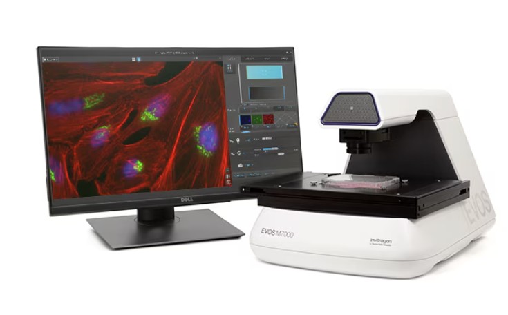
The SCIF operates an EVOS M7000 inverted fluorescent microscope with stage automation. The microscope is capable of 4 channels of fluorescence, plus brightfield. The stage automation enables x-y stitching and z-stacks. Additionally, there is a color camera useful for histological evaluation.
Objectives:
4x Objective: EVOS fluorite, LWD, phase-contrast, 0.13NA/10.58WD - long working distance, good to image through thicker vessels
10x Objective: EVOS fluorite, LWD, phase-contrast, 0.30NA/7.13WD - long working distance, good to image through thicker vessels
20x Objective: EVOS fluorite, LWD, phase-contrast, 0.45NA/6.12WD - long working distance, good to image through thicker vessels
40x Objective: EVOS fluorite, coverslip-corrected - short working distance, best to image through cover slip
60x Objective: Olympus™ fluorite, 0.90NA/0.2WD, correction collar (0.11–0.23 mm) - short working distance, best to image through cover slip
Filter Cubes:
DAPI - Ex: 357/44 Em: 447/60
GFP - Ex: 482/25 Em: 524/24
TexasRed - Ex: 585/29 Em: 628/32
Cy5 - Ex: 635/18 Em: 692/40
RFP (Requires filter change, ask staff for guidance) - Ex: 542/20 Em: 593/40
Histology Services
***Please note that as of 10/1/25 histology services provided by SCIF, such as tissue processing, embedding, and microtomy have been transfered to the RESTORE core facility***
